Your shopping cart is empty!
MENU
Your shopping cart is empty!
 Electrophoresis is one of the simplest yet most sensitive analytical tools used in separating proteins, nucleic acids and other biological molecules in sample solutions. However, did you know that there are about eight types of electrophoresis and that each one of these techniques can give you a unique and valuable piece of information about your target protein? If you want to learn more about these different types of electrophoretic protocols, then let’s get started!
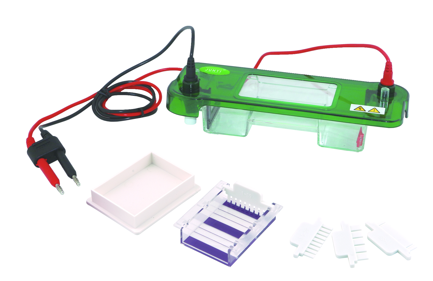
Routine electrophoresis is the traditional and most widely used clinical laboratory technique for separating proteins and nucleic acids. This technique is usually performed on a rectangular-shaped slab gel and is also called “zone electrophoresis” since it can accommodate several specimens and controls on one gel, and can be used to separate solutes in a single run. It can also be used in separating CSF and urine proteins, isoenzymes, lipoproteins, and hemoglobin.
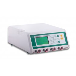
High-resolution electrophoresis (HRE) is nothing but routine electrophoresis using a high voltage. This technique usually comes highly recommended if you require higher protein resolution (e.g. separation of CSF proteins for the detection of multiple sclerosis, separation of light chains in urine for early detection of multiple myeloma, etc.). Since increasing the voltage also increases the heat produced, HRE includes a cooling instrumentation to prevent denaturation of proteins and the drying out of gel and other components.
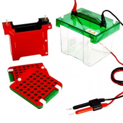
Electrophoresis by acrylamide (also known as PAGE) is usually used to separate proteins based on molecular size and charge-to-mass ratio. With the help of vertical slabs or gel incorporated in vertical rods or cylinders, researchers can separate DNA of 100 base pairs or less and analyze individual proteins in a serum (e.g. genetic variants, isoenzymes). Aside from its simplicity and speed of separation, researchers love PAGE since the gels are stable over a wide range of pH and temperature, and gels of different pore size can be formed.
Capillary electrophoresis is performed in submillimeter diameter capillaries (i.e. extremely thin, fused silica capillary tubes with internal an diameter of 25 to 100 mm) and combines electrophoresis and high-performance liquid chromatography to facilitate analyte separation. A good number of researchers prefer using CE because it only requires a small amount of sample, is extremely efficient, produces rapid results, and can be easily automated.
If you want to separate amphoteric compounds (e.g. proteins) with higher resolution, then you need to use this protocol. IEF uses chemical infused gels to create a pH gradient across the surface of the gel and applies an extremely high voltage to facilitate the migration of protein molecules up to the point where their net charge is zero (isoelectric point). Some of the advantages of using IEF include the ease of procedure (i.e. placement of sample application is not important since the protein will always end up in a position corresponding to its pl) and its high resolution.
Generally, IFE is used in the detection of monoclonal immunoglobulin gammopathies or monoclonal expansions of a single, non-functional antibody such as IgA, IgG and IgM, the presence of which may indicate conditions such as multiple myeloma or Waldenstrom’s macroglobulinemia. It can also be used in studying protein antigens and their split products.
You cannot separate large DNA molecules >50 kilobases (kb) by using AGE or PAGE in routine electrophoresis systems because the gel pore sizes are simply too small to allow for their migration. However, you can use pulsed field gel electrophoresis (PFGE) to facilitate the successful fractionation of large DNA molecules (up to 10 Mb).
PFGE effectively separates DNA fragments by applying an electric current that constantly changes direction onto the gel matrix. By alternating the positive and negative electrodes in cycles during electrophoresis, the DNA molecules are forced to reorient themselves and eventually break down into smaller fragments. PFGE is commonly used in genotyping or genetic fingerprinting and is considered as the gold standard in bacterial subtyping due to its simplicity and reproducibility.
However, this protocol is extremely time-consuming and requires a high level of skills. Additionally, the results may be difficult to interpret since the fragments are separated according to their size (i.e. separation not based on sequence) and fragments of the same size may not come from the same part of the chromosome.
In two-dimensional electrophoresis, the sample is separated using two distinct separation techniques (e.g. IEF followed by PAGE or AGE) and identified in two dimensions oriented at right angles to each other. Since the resulting bands are further resolved with the second electrophoresis, there is a great possibility that you will gain more information from your sample.
Two-dimensional electrophoresis is highly specialized and is commonly used in proteomics and genetics research. While it can analyze thousands of proteins in a single run, this technique requires a substantial amount of starting sample, has limited reproducibility, and is low throughput. Additionally, this technique works only with medium to large sized biomolecules and does not produce precise measurements.
")
")
")
")

")
")
Leave a Comment