Your shopping cart is empty!
MENU
- +
-
Categories+
- Blog +
- Contact Us +
- Login +
- Register +
 An Introduction to Electrophoresis
Electrophoresis is a simple, sensitive laboratory technique essential for the separation and isolation of proteins, nucleic acids and other biological molecules in sample solutions. Based on their size and charge, the molecules will travel through the gel at various directions and speeds and separate from one another.
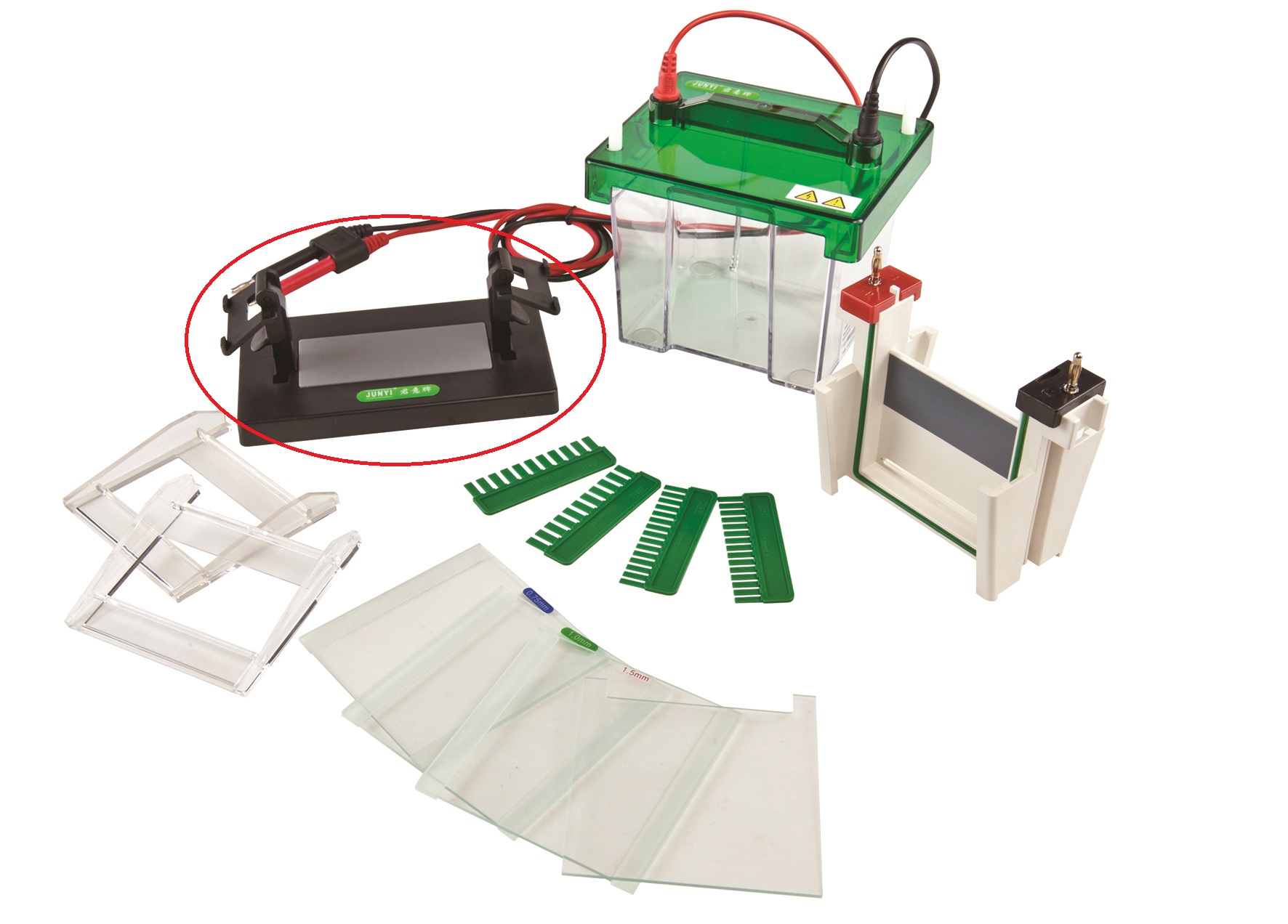
In electrophoresis, the molecules are separated by an electrical field through a gel apparatus, which contains a cathode on one end, an anode on the other, and a platform that holds a porous gel matrix in the middle (such as agarose or acrylamide gel). Then, a buffer-filled box is added to create a charge gradient upon the application of an electric current. When a charge is applied, the buffer prevents the gel from overheating and keeps it cool.
Due to a uniformly net-negative charge, proteins and nucleic acids naturally migrate towards the positive electrode. Since smaller molecules easily move through the pores of the gel matrix, they migrate faster than larger molecules. Once electrophoresis is complete, researchers will observe proteins and nucleic acids, separated based upon molecular weights.
There are ten main types of electrophoresis, including protein electrophoresis, vertical electrophoresis and polyacrylamide electrophoresis.
As the name suggests, protein electrophoresis involves proteins, which are positively charged. Due to a lack of negative charge, they cannot be separated by the application of an electric field. To overcome the issue, detergent sodium dodecyl sulfate is added to the protein solution, which separates proteins using gel electrophoresis.
Protein electrophoresis allows the proteins to unfold into a linear shape, while covering them with a negatively charged coat, which allows proteins to migrate towards the positively charged end of a gel and be separated by molecular weight.
More complex than horizontal systems, vertical electrophoresis is ideal for proteins. It utilizes a discontinuous buffer system, where the top chamber contains a cathode and the bottom chamber an anode. The buffer can only flow through the gel, allowing for precise control of voltage gradients during separation. When combined with the smaller pore size of the acrylamide gel, greater separation and resolution is achieved.
As one of the most common laboratory methods, polyacrylamide electrophoresis separates biological macromolecules, according to their electrophoretic mobility, which is defined as a function of length, conformation and charge of the molecule. Macromolecules, such as proteins or nucleic acids, may run in either a native state or in denatured forms.
In polyacrylamide electrophoresis, to separate molecules based on length, samples are run in denaturing conditions. As a result, negatively charged proteins will migrate towards the positive electrode and will be fractioned by approximate size during electrophoresis.
BT Lab Systems offers a variety of units suitable for electrophoresis. For more information about our electrophoresis products and warranties, visit our website today.
Leave a Comment