Western blotting (also called protein blotting and/or immunoblotting) is a widely accepted analytical technique for the detection and characterization of proteins in a given sample. To accomplish this task, western blotting makes use of two processes - gel electrophoresis and protein blotting and testing. Here’s how it works.
The Principles of Western Blotting
Gel Electrophoresis
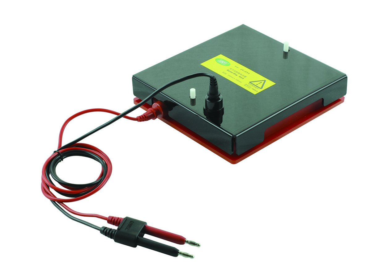
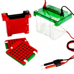
After suitable samples (i.e., cell lysates, tissue samples and/or purified or semi-purified extracts) have been prepared, small volumes of the sample (about 5 to 20 µl) are dissolved in buffer containing sodium dodecyl sulfate (SDS) and loaded into a commercially available polyacrylamide gel. The presence of SDS serves to unfold the proteins (by breaking the hydrogen bonds within and between the protein molecules and removing any secondary and tertiary structures present in the sample) and impart a uniformly negative charge to the molecules.
Since the addition of SDS equalizes the charge-to-mass ratio and imparts a new negative charge, which is significantly greater than the original charge of the protein molecules, the application of an electric field will cause them to move in the direction of the positive electrode based primarily on their size. This means that their native charge and structure will not have any effect on their electrophoretic mobility.
When running the gel, care should be taken to ensure that the proper voltage is maintained throughout the procedure since high voltage may cause overheating and distort the resulting bands.
There are a number of reasons why polyacrylamide gel is ideally used for this purpose.
- It has a neutrally charged nature, is thermo-stable and chemically inert (i.e. will not react to samples).
- You can use different buffers in your experiment to manipulate resolution and run time.
- PA gel is synthetic so you can expect to get more consistent results between batches.
- Pore size can be manipulated to increase and/or decrease molecular sieving.
- The percentage and thickness of the gel affect the transfer of protein to the blotting phase so if you want to improve your results, consider using a lower percentage of acrylamide or a thinner gel.
However, since acrylamide is a potent neurotoxin, care should be taken when working with the solution.
Western Blotting in 5 Steps
Western blotting is performed in five steps – transfer, blocking, incubation with primary antibody, incubation with secondary antibody, and protein detection and analysis.
- Transfer. First, proteins are taken from the gel to a nitrocellulose or PVDF membrane through the application of an electric field. This process can occur under standard wet or semi-dry conditions. Semi-dry transfers are quicker and easier than standard wet transfers, but they are also significantly more expensive and may produce patchy or uneven blots.
- Blocking. After transferring the proteins to the membrane, blocking buffers (e.g., BSA, 3%-5% non-fat dry milk, casein, gelatin, and Tween-20) are used to block unreacted areas on the blot (i.e., areas that do not contain protein). This step reduces overall background signal and prevents non-specific binding of the antibody to the blotting membrane.
- Primary Antibody Incubation. After blocking, the membrane is rinsed in wash buffer and incubated at room temperature for a few hours or overnight at 4ºC in a dilute antibody solution (monoclonal or polyclonal) specific to the target protein. To remove unbound primary antibody, wash the membrane vigorously in TBS five times for five minutes each.
- Secondary Antibody Incubation. The membrane can now be exposed to specific enzyme conjugated secondary antibody (e.g., anti-mouse and anti-rabbit immune globulin) to bind with the primary antibody.
- Protein Detection and Analysis. After a series of wash steps, the blot is finally ready for protein detection. Some of the detection methods used include electrochemiluminescence (ECL), colorimetric, fluorescence and radioactive detection.
Western blots are widely used in protein laboratories worldwide since it is fast and easy to implement, uses simple equipment and inexpensive reagents, and produces results that are clear and easy to interpret.
Related Blogs


Leave a Comment